Monday, September 30, 2013

A&P Lecture 1.2: Cytoplasm

This is an in depth  explanation of the Cytoplasm as a part of the cell. This lecture note is linked to A&P Lecture 1: The Cell


ANATOMY AND PHYSIOLOGY LECTURE NOTE 1.2
CYTOPLASM, CYTOSOL, CYTOSKELETON, 
AND CYTOPLASMIC INCLUSION


CYTOPLASM
Cytoplasm, the cellular material outside the nucleus but inside the plasma membrane, is about half cytosol and half organelles.

CYTOSOL
Cytosol consists of a fluid portion, a cytoskeleton, and cytoplasmic inclusions. The fluid portion of cytosol is a solution with dissolved ions and molecules and a colloid with suspended molecules, especially proteins. Many of these proteins are enzymes that catalyze the breakdown of molecules for energy or the synthesis of sugars, fatty acids, nucleotides, amino acids, and other molecules.

CYTOSKELETON
The cytoskeleton supports the cell and holds the nucleus andother organelles in place. It is also responsible for cell movements,such as changes in cell shape and the movement of cell organelles. The cytoskeleton consists of three groups of proteins: microtubules, actin filaments, and intermediate filaments ( figure 3.27 ).
Microtubules are hollow tubules composed primarily of protein units called tubulin. The microtubules are about 25 nanometers (nm) in diameter, with walls about 5 nm thick. 

Microtubules vary in length but are normally several micrometers (μm) long. Microtubules play a variety of roles within cells. They help provide  support and structure to the cytoplasm of the cell, much like an internal scaffolding. They are involved in the process of cell division and in the transport of intracellular materials, and they form essential components of certain cell organelles, such as centrioles, spindle fibers, cilia, and flagella.

Actin filaments, or microfilaments, are small fibrils about 8 nm in diameter that form bundles, sheets, or networks in the cytoplasm of cells. These filaments have a spiderweb-like appearance within the cell. Actin filaments provide structure to the cytoplasm and mechanical support for microvilli. Actin filaments support the plasma membrane and define the shape of the cell. Changes in cell shape involve the breakdown and reconstruction of actin filaments. These changes in shape allow some cells to move about. Muscle cells contain a large number of highly organized actin filaments, which are responsible for the muscle’s contractile capabilities.

Intermediate filaments are protein fibers about 10 nm in diameter. They provide mechanical strength to cells. For example, intermediate filaments support the extensions of nerve cells, which have a very small diameter but can be a meter in length. 

CYTOPLASMIC INCLUSION
The cytosol also contains cytoplasmic inclusions, which are aggregates of chemicals either produced by the cell or taken in by the cell.For example, lipid droplets or glycogen granules store energy-rich molecules; hemoglobin in red blood cells transports oxygen; melanin is a pigment that colors the skin, hair, and eyes; and lipochromes are pigments that increase in amount with age. Dust, minerals, and dyes can also accumulate in the cytoplasm.

A&P Lecture 1.3: Nucleus

This lecture note on Anatomy and Physiology is linked to A&P Lecture 1: The Cell to further explain the physiology of the Nucleus.


ANATOMY AND PHYSIOLOGY LECTURE 1.3
NUCLEUS


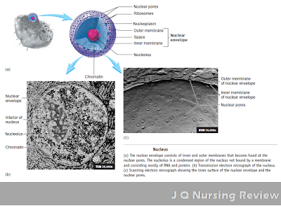
PARTS OF A NUCLEUS


The nucleus is a large, membrane-bound structure usually located near the center of the cell. It may be spherical, elongated, or lobed, depending on the cell type. All cells of the body have a nucleus at some point in their life cycle, although some cells, such as red blood cells, lose their nuclei as they develop. Other cells, such as skeletal muscle cells and certain bone cells, called osteoclasts, contain more than one nucleus.

The nucleus consists of nucleoplasm surrounded by a nuclear envelope composed of two membranes separated by a space. At many points on the surface of the nuclear envelope, the inner and outer membranes fuse to form porelike structures called nuclear pores. Molecules move between the nucleus and the cytoplasm through these nuclear pores.

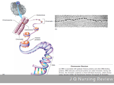
Deoxyribonucleic acid (DNA) is mostly found within the nucleus , although small amounts of DNA are also found within mitochondria. Nuclear DNA and associated proteins are organized into discrete structures called chromosomes. The proteins include histones, which are important for the structural organization of DNA, and other proteins that regulate DNA function. During most of the life cycle of a cell, the chromosomes are dispersed throughout the nucleus as delicate filaments referred to as chromatin. During cell division, the dispersed chromatin becomes densely coiled, forming compact chromosomes. At the beginning of cell division, each chromosome consists of two chromatids , which are attached at a single point called the centromere. The kinetochore, a protein structure within the centromere, provides a point of attachment for microtubules during cell division.


Chromosomal Structure


DNA determines the structural and functional characteristics of the cell by specifying the structure of proteins. Proteins form many structural components of the cell and all the enzymes, which regulate most chemical reactions in the cell. DNA establishes the structure of proteins by specifying the sequence of their amino acids. DNA is a large molecule that does not leave the nucleus but functions by means of an intermediate, ribonucleic acid (RNA) , which can leave the nucleus through nuclear pores. DNA determines the structure of messenger RNA (mRNA), ribosomal RNA (rRNA), and transfer RNA (tRNA) (all described in more detail on p. 86). A sequence of nucleotides in a DNA molecule that specifies the structure of a protein or RNA molecule is called a gene.

Because mRNA synthesis occurs within the nucleus, cells without nuclei accomplish protein synthesis only as long as the mRNA produced before the nucleus degenerates remains functional. The nuclei of developing red blood cells are expelled from the cells before the red blood cells enter the blood, where they survive without a nucleus for about 120 days. In comparison, many cells with nuclei, such as nerve and skeletal muscle cells,
survive as long as the person survives. 

A nucleolus is a somewhat rounded, dense region within the nucleus that lacks a surrounding membrane . Usually, one nucleolus exists per nucleus, but several nucleoli may also be seen in the nuclei of rapidly dividing cells. The nucleolus incorporates portions of 10 chromosomes (5 pairs), called nucleolar organizer regions. These regions containDNA from which rRNA is produced. Within the nucleolus, the subunits of ribosomes are manufactured

A&P Lecture 1.1: Cell Membrane

This lecture note on Anatomy and Physiology is linked to A&P Lecture 1: The Cell to further explain the physiology of the Cell Membrane. 


ANATOMY AND PHYSIOLOGY LECTURE NOTE 1.1
CELL MEMBRANE


The outermost component of the cell is the plasma membrane. It is a boundary that separates the substances inside the cell, which are intracellular , from substances outside the cell, which are extracellular.It determines what moves into and out of cells. As a result, the intracellular contents of cells is different from the extracellular environment. The membrane encloses and supports the cell contents. It attaches cells to the extracellular environment or to other cells. The cells’ ability to recognize and communicate with each other takes place through the plasma membrane.

The regulation of ion movement by cells results in a charge difference across the plasma membrane called the membrane potential. The outside of the plasma membrane is positively charged, compared with the inside, because there are more positively charged ions immediately on the outside of the plasma membrane and more negatively charged ions and proteins inside. This concept is paramount in understanding the Sodium Potassium Pump that controls the electrophysiology of the heart, transmission of nerve impulses and contraction of muscles.

The plasma membrane consists of 45%–50% lipids, 45%– 50% proteins, and 4%–8% carbohydrates ( figure 3.2 ). The carbohydrates combine with lipids to form glycolipids and with proteins to form glycoproteins. The glycocalyx (glı¯-ko¯-ka¯ liks) is the collection of glycolipids, glycoproteins, and carbohydrates on the outer surface of the plasma membrane. The glycocalyx also contains molecules absorbed from the extracellular environment, so there is often no precise boundary where the plasma membrane ends and the extracellular environment begins.

Indepth Medical Physiology

The membrane that surrounds cells is a notable structure. It is made up of lipids and proteins and is semipermeable meaning- it allows some substances to pass through it and excluding others. However, its permeability can also be varied because it contains numerous regulated ion channels and other transport proteins that can change the amounts of substances moving across it. It is generally referred to as the plasma membrane. The nucleus and other organelles in the cell are bound by similar membranous structures.

Although the chemical structures of membranes and their properties vary considerably from one location to another, they have certain common features. They are generally about 7.5 nm thick. The major lipids are phospholipids such as phosphatidylcholine, phosphotidylserine, and phosphatidylethanolamine. The shape of the phospholipid molecule reflects its solubility properties: the “head” end of the molecule contains the phosphate portion and is relatively soluble in water  and the “tail” ends are relatively insoluble. The possession of both hydrophilic and hydrophobic properties makes the lipid an amphipathic molecule. In the membrane, the hydrophilic ends of the molecules are exposed to the aqueous environment that bathes the exterior of the cells and the aqueous cytoplasm; the hydrophobic ends meet in the water-poor interior of the membrane. In prokaryotes, the membranes are relatively simple, but in eukaryotes, cell membranes contain various glycosphingolipids, sphingomyelin, and cholesterol in addition to phospholipids and phosphatidylcholine.

Many different proteins are embedded in the membrane. They exist as separate globular units and many pass through or are embedded in one leaflet of the membrane- integral proteins, whereas others- peripheral proteins, are associated with the inside or outside of the membrane. The amount of protein varies significantly with the function of the membrane but makes up on average 50% of the mass of the membrane; that is, there is about one protein molecule per 50 of the much smaller phospholipid molecules.

The proteins in the membrane carry out many functions. Some are cell adhesion molecules that anchor cells to their neighbors or to basal laminas. Some proteins function as pumps, actively transporting ions across the membrane. Other proteins function as carriers, transporting substances down electrochemical gradients by facilitated diffusion. Still others are ion channels, which, when activated, permit the passage of ions into or out of the cell. The role of the pumps, carriers, and ion channels in transport across the cell membrane is discussed below. Proteins in another group function as receptors that bind ligands or messenger molecules, initiating physiologic changes inside the cell. Proteins also function as enzymes, catalyzing reactions at the surfaces of the membrane. 

The uncharged, hydrophobic portions of the proteins are usually located in the interior of the membrane, whereas the charged, hydrophilic portions are located on the surfaces. Peripheral proteins are attached to the surfaces of the membrane in various ways. One common way is attachment to glycosylated forms of phosphatidylinositol. Proteins held by these glycosylphosphatidylinositol (GPI) anchors include enzymes such as alkaline phosphatase, various antigens, a number of CAMs, and three proteins that combat cell lysis by complement. Over 45 GPI-linked cell surface proteins have now been described in humans. Other proteins are lipidated, that is, they have specific lipids attached to them. Proteins may be myristoylated, palmitoylated, or prenylated (ie, attached to geranylgeranyl or farnesyl groups).

The protein structure—and particularly the enzyme content—of biologic membranes varies not only from cell to cell, but also within the same cell. For example, some of the enzymes embedded in cell membranes are different from those in mitochondrial membranes. In epithelial cells, the enzymes in the cell membrane on the mucosal surface differ from those in the cell membrane on the basal and lateral margins of the cells; that is, the cells are polarized. Such polarization makes directional transport across epithelia possible. Th e membranes are dynamic structures, and their constituents are being constantly renewed at different rates. Some proteins are anchored to the cytoskeleton, but others move laterally in the membrane.

Underlying most cells is a thin, “fuzzy” layer plus some fibrils that collectively make up the basement membrane or, more properly, the basal lamina. Th e basal lamina and, more generally, the extracellular matrix are made up of many proteins that hold cells together, regulate their development, and determine their growth. These include collagens, laminins, fibronectin, tenascin, and various proteoglycans.

Case Study #0001: Orthostatic Hypotension

The Case Study Series of JQ Nursing Review presents a real life patient situation to encourage you to become an active learner as you read. This will also develop your critical thinking skills. Case Study Questions will challenge you to use your understanding of new concepts and help you integrate your knowledge to solve a clinical problem.

Case Study #0001: 
Orthostatic Hypotension


Scenario
As a nurse, you are assigned at the Emergency Room of the hospital to provide rapid assessment and intervention to various clients.

 In your shift a 72-year old woman was accompanied by her son presented herself at the emergency room. You asked what brings her to the hospital and you've found out that for 3 days, she have had  fever and chills and stayed mostly in bed. On rising to go to the bathroom, she felt dizzy, fainted, and fell to the floor. She regained consciousness immediately and called her son.

Upon further assessment and evaluation by the health team, a diagnosis of Orthostatic Hypotension was made.

Clinical Questions
a. Describe the normal response to a decrease in blood pressure on standing.
b. What happened to the client's heart rate just before she fainted? Why did the client faint?
c. How did the client's fainting and falling to the floor assist in establishing homeostasis (assuming she was not injured)?

A&P Lecture 3: Homeostasis

"You need to know the normal before you can determine the abnormal.

Knowledge in Anatomy and Physiology is paramount before tackling advance Nursing Courses. It is the reason why Anatomy and Physiology class is taken first before professional courses like Pathophysiology, Medical Surgical Nursing, Maternal and Child Nursing and Psychiatric Nursing.Anatomy and Physiology is a good foundation when you already try to study illnesses. This A&P Lecture Series is designed to help students develop a solid understanding of the concepts of anatomy and physiology and to use this knowledge to solve problems.


ANATOMY AND PHYSIOLOGY LECTURE 2
HOMEOSTASIS


Homeostasis is defined as the body's continuous maintenance of a relatively constant environment within the body. Homeostasis is also defined as the condition of equilibrium or balance in the body’s internal environment beacuse to the constant interaction of the different regulatory processes in the body. Homeostasis is a dynamic condition. In response to changing conditions, the body’s equilibrium can shift among points in a narrow range that is compatible with maintaining life.

Such Homeostatic mechanisms, such as sweating or shivering, normally maintain body temperature near an ideal normal value, or set point. It is important to note that these mechanisms are not able to maintain body temperature precisely at the set point. Instead, body temperature increases and decreases slightly around the set point to produce a normal range of values. As long as body temperature remains within this normal range, homeostasis is maintained.

The organ systems help control the body ’s internal environment so that it remains relatively constant. For  example, the digestive, respiratory, circulatory, and urinary systems function together so that each cell in the body receives adequate oxygen and nutrients and so that waste products do not accumulate to a toxic level. If the fluid surrounding cells deviates from homeostasis, the cells do not function normally and can even die. Disease disrupts homeostasis and sometimes results in death.


Body Fluids and Electrolytes and Acid Base Balance
An integral aspect of homeostasis is maintaining the volume and composition of body fluids, dilute, watery solutions containing dissolved chemicals that are found inside cells as well as surrounding them. This s a pivotal concept in understanding the mechanism of fluid and electrolyte movement inside the body.

A good example s when the inside of a cell has more fluid in it compared to its surrounding environment, the body will activate several mechanism to equalize the pressure gradient between these two compartments.
     ***This concept will be further explained in the upcoming lecture notes on Fluid and Electrolytes and Acid Base Balace.

Control
The body has many regulating systems that can usually bring the internal environment back into balance. Most often, the nervous system and the endocrine system, working together or independently, provide the needed corrective measures. Both means of regulation, however, work toward the same end, usually through negative feedback systems.

Feedback System
A feedback system or feedback loop is a cycle of events in which the status of a body condition is monitored, evaluated, changed, remonitored, reevaluated, and so on.

Parts
1. A receptor is a body structure that monitors changes in a controlled condition and sends input to a control center.
2. A control center in the body, for example, the brain, sets the range of values within which a controlled condition should be maintained, evaluates the input it receives from receptors, and generates output commands when they are needed.
3. An effector is a body structure that receives output from the control center and produces a response or effect that changes the controlled condition.

Negative Feedback
Most systems of the body are regulated by negative-feedback mechanisms, which maintain homeostasis. Negative means that any deviation from the set point is made smaller or is resisted.


Positive Feedback
Positive-feedback responses are not homeostatic and are rare in healthy individuals. Positive implies that, when a deviation from a normal value occurs, the system ’s response is to make the deviation even greater.


Sunday, September 29, 2013

A&P Lecture 1: The Cell

"You need to know the normal before you can determine the abnormal.

Knowledge in Anatomy and Physiology is paramount before tackling advance Nursing Courses. It is the reason why Anatomy and Physiology class is taken first before professional courses like Pathophysiology, Medical Surgical Nursing, Maternal and Child Nursing and Psychiatric Nursing.Anatomy and Physiology is a good foundation when you already try to study illnesses. This A&P Lecture Series is designed to help students develop a solid understanding of the concepts of anatomy and physiology and to use this knowledge to solve problems.


ANATOMY AND PHYSIOLOGY LECTURE 1
THE CELL


The Cell is the basic unit of life. Cells are the smallest parts of an organism, such as a human, that have the characteristics of life. Although cells may have quite different structures and functions, they share several characteristics which includes:

  • Cell metabolism and energy use
  • Synthesis of molecules
  • Communication
  • Reproduction and inheritance

Plasma Membrane

Structure: Lipid bilayer composed of phospholipids and cholesterol with proteins that extend across or are embedded in either surface of the lipid bilayer
Function: Outer boundary of cells that controls entry and exit of substances; receptor molecules function in intercellular communication; marker molecules enable cells to recognize one another.

***Read more about the Plasma Membrane here

Cytoplasm
Fluid Part
Structure: Water with dissolved ions and molecules; colloid with suspended proteins
Function:Contains enzymes that catalyze decomposition and synthesis reactions; ATP is produced in glycolysis reactions

Cytoskeleton/ Microtubules
Structure: Hollow cylinders composed of the protein tubulin; 25 nm in diameter
Function: Support the cytoplasm and form centrioles, spindle fibers, cilia, and flagella; responsible for movement of structures in the cell

Actin Filaments
Structure: Small fibrils of the protein actin; 8 nm in diameter
Function: Provide structural support to cells, support microvilli, responsible for cell movements

Intermediate Filaments
Structure:Protein fibers; 10 nm in diameter
Function: Provide structural support to cells

Cytoplasmic Inclusions
Structure:Aggregates of molecules manufactured or ingested by the cell; may be membrane-bound
Function: Function depends on the molecules: energy storage (lipids, glycogen), oxygen transport (hemoglobin), skin color (melanin), and others


*** Read More about the Cytosol and its components here

Organelles, Nucleus
Nuclear envelope
Structure:Double membrane enclosing the nucleus; the outer membrane is continuous with the endoplasmic reticulum; nuclear pores extend through the nuclear envelope
Function: Separates nucleus from cytoplasm and regulates movement
of materials into and out of the nucleus

Chromatin
Structure:Dispersed, thin strands of DNA, histones, and other proteins; condenses to form chromosomes during cell division
Function: DNA regulates protein (e.g., enzyme) synthesis and therefore the chemical reactions of the cell; DNA is the genetic, or hereditary, material

Nucleolus
Structure: One or more dense bodies consisting of ribosomal RNA and proteins
Function: Assembly site of large and small ribosomal subunits

***Read more about the Nucleus and it's components here

Cytoplasmic Organelles
Ribosome
Structure:Ribosomal RNA and proteins form large and small subunits; attached to endoplasmic reticulum or free ribosomes are distributed throughout the cytoplasm
Function: Site of protein synthesis
 ***Read more about Ribosome here


Rough endoplasmic reticulum
Structure: Membranous tubules and flattened sacs with attached ribosomes
Function: Protein synthesis and transport to Golgi apparatus
*** Read more about the Endoplasmic Reticulum here


Smooth endoplasmic reticulum
Structure: Membranous tubules and flattened sacs with no attached ribosomes
Function: Manufactures lipids and carbohydrates; detoxifies harmful chemicals; stores calcium
*** Read more about the Endoplasmic Reticulum here

Golgi apparatus
Structure: Flattened membrane sacs stacked on each other
Function: Modifies, packages, and distributes proteins and lipids for secretion or internal use
***Read more about the Golgi apparatus here

Secretory vesicle
Structure: Membrane-bound sac pinched off Golgi apparatus
Function: Carries proteins and lipids to cell surface for secretion
***Read more about the Secretory vesicle here

Lysosome
Structure: Membrane-bound vesicle pinched off Golgi apparatus
Function: Contains digestive enzymes
***Read more about the Lysosomes  here


Peroxisome
Structure: Membrane-bound vesicle
Function: One site of lipid and amino acid degradation; breaks down hydrogen peroxide
***Read more about Peroxisome here


Proteasomes
Structure: Tubelike protein complexes in the cytoplasm
Function: Break down proteins in the cytoplasm
***Read more about Proteasomes here


Mitochondria
Structure: Spherical, rod-shaped, or threadlike structures; enclosed by double membrane; inner membrane forms projections called cristae
Function: Major site of ATP synthesis when oxygen is available
***Read more about the Mitochondria here


Centrioles
Structure: Pair of cylindrical organelles in the centrosome, consisting of triplets of parallel microtubules 
Function: Centers for microtubule formation; determine cell polarity during cell division; form the basal bodies of cilia and flagella
***Read more about the Centriole here

Spindle fibers
Structure:Microtubules extending from the centrosome to chromosomes and other parts of the cell (i.e., aster fibers)
Function: Assist in the separation of chromosomes during cell division
***Read more about the Spindle fiber here

Cilia
Structure: Extensions of the plasma membrane containing doublets of parallel microtubules; 10 μm in length
Function: Move materials over the surface of cells
***Read more about the Cilia here

Flagellum
Structure: Extension of the plasma membrane containing doublets of parallel microtubules; 55 μm in length
Function: In humans, responsible for movement of spermatozoa
***Read more about the Flagellum here

Microvilli
Structure: Extension of the plasma membrane containing microfilaments
Function: Increase surface area of the plasma membrane for absorption and secretion; modified to form sensory receptors
***Read more about the Microvillli here

About JQ Nursing Review

Nursing, a noble profession is indeed have become a very popular career of choice for up-coming college students. With the continuous development of science and technology as well as advancements in the field of health care many illnesses are being treated with various modalities in diverse health settings thus increasing the need for nurses to as part of a team to cater for the needs of those who seek for medical help. Moreover, new laws and regulations are enacted to provide affordable health services with new health insurances thus making health care services available, affordable and accessible to many people especially the underprivileged. The improved health seeking behaviors seen in people also increases the demand for a number of nurses to serve and provide quality care to these health consumers.

With this advent, authorities in the Nursing Profession had continually developed new curriculum to train future nurses to be nurses who are competent, compassionate and responsive to the needs of every client in various health care settings. To regulate the practice of nursing and to ensure optimum level of proficiency be observed in the profession of nursing, nursing authorities/ agencies have included an examination for nursing before fully entering the profession and be able to care to clients. 

JQ Nursing Review is an complete and comprehensive online nursing review experience that will cater to every needs to nurses-to-be in studying concepts in learned in nursing school as well as a preparation for the exams- may it be a quiz, final term exam or the NCLEX-RN, NLE, HAAD Examination. 

Topics are carefully written to be able to provide quality materials to nurses and nurses-to-be to deliver them in the simplest way possible for an easy grasp of the topic.

Lectures are presented in various ways to provide a wide range of learning experience- traditional and case analysis method.


Moreover, to fully develop your skills and attitude for the exam, JQ Nursing Review also provides quality test questions from trusted resources to fully maximize your review experience.


What's in the Review Site?

  • FREE Lecture Notes in Nursing from reliable and well known sources.
  • FREE Nursing Drills
  • FREE Study Guide
  • FREE Books
  • FREE NCLEX-RN, NLE, HAAD Exam Test Questions


    Tuesday, September 17, 2013

    Nursing Leader: Linda Richards

    This is a series of post regarding notable nursing leaders who have given many contribution to nursing and their influence uplifted the nursing profession.


    Nursing Leaders
    Florence Nightingale, Clara Barton, Lillian Wald, Lavinia Dock, Margaret Sanger, and Mary Breckinridge are among the leaders who have made notable contributions both to nursing's history and to women's history. These women were all politically astute pioneers. Their skills at influencing others and bringing about change remain models for political nurse activists today. Contemporary nursing leaders, such as Virginia Henderson, who created a modern worldwide definition of nursing, and Martha Rogers, a catalyst for theory  development.

    Richards (1841-1930)
    Linda Richards  was America's first trained nurse. She graduated from the New England Hospital for Women and Children in 1873. Richards is known for introducing nurse's notes and doctor's orders. She also initiated the practice of nurses wearing uniforms (American Nurses Association, 2006a). She is credited for her pioneer work in psychiatric and industrial nursing

    Born: Linda Richards was born on July 27, 1841, the youngest daughter of Sanford Richards, an itinerant preacher, and his wife, Betsy Sinclair Richards.

    After ten years as a schoolteacher, began working as a nurse at Boston City Hospital in 1870. She enrolled for training in 1872 at the New England Hospital for Women and Children, run by female physicians, for a one-year course based on the principles established by Florence Nightingale. Linda received her diploma on September 1, 1873, and went to work as night supervisor at Bellevue Hospital in New York.

    After attending Florence Nightingale's training school at St. Thomas Hospital in England in 1877, became superintendent of a new training school at Boston City Hospital, which officially opened in 1878.

    Worked in Japan for five years beginning in 1886 to start a training school for nurses. Back in the United States, worked as a visiting nurse and helped train nurses to work with the mentally ill.

    She retired in 1911 at age 70 when she wrote her autobiography, Reminiscences of Linda Richards. She suffered a severe stroke in 1923 and lived the remainder of her life at the New England Hospital for Women and Children where she had done her first training. She died on April 16, 1930 in Boston.